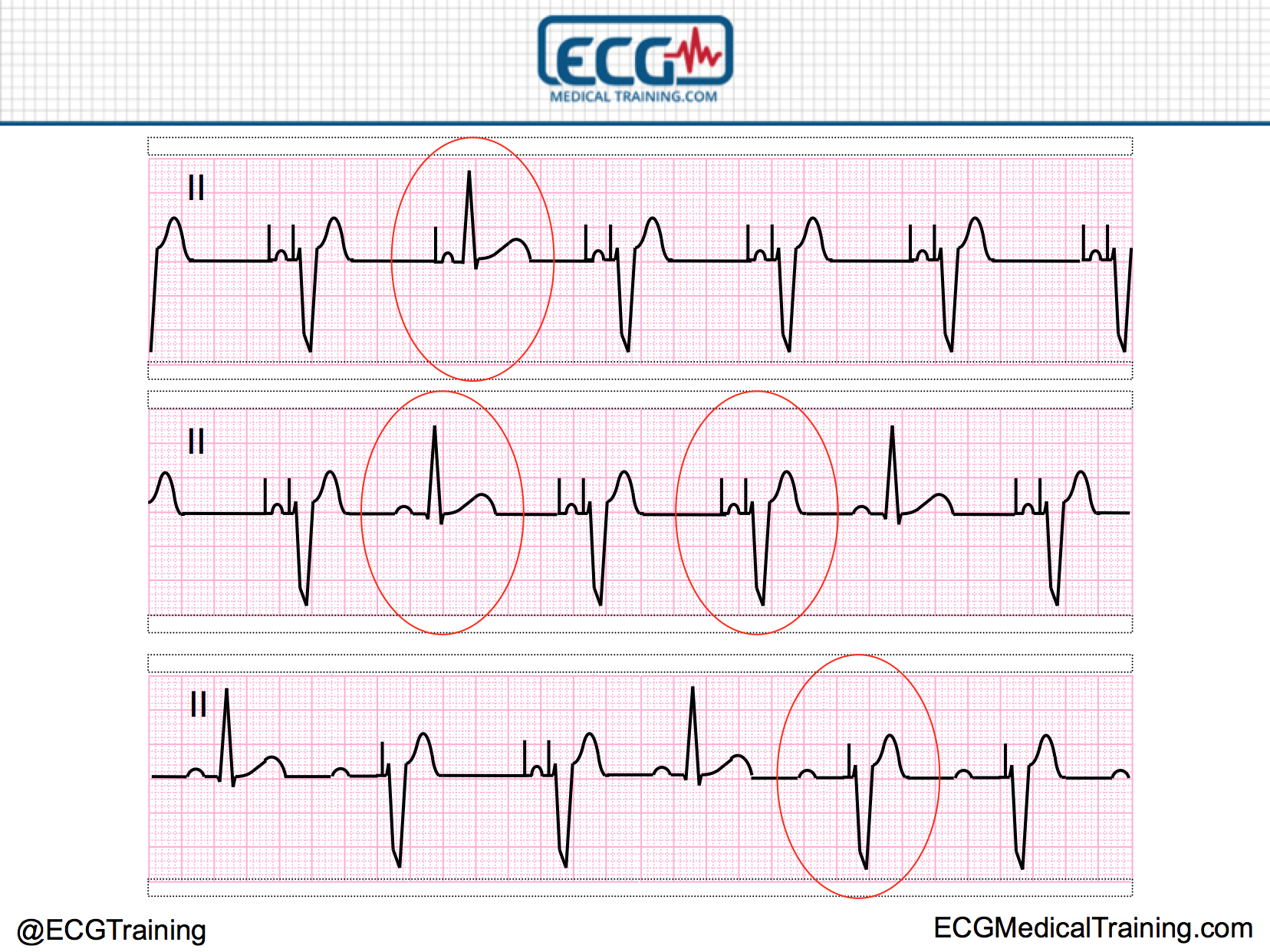

Again, the cardiac rhythm is dependent on the patient’s underlying cardiac rhythm. There are a few million pacemaker patients worldwide, with hundreds of thousands. Pacer spikes can be seen on the ECG, but without an associated P wave or QRS complex. An accurate diagnosis of pacing system malfunction (s) requires knowledge of different modes, timing cycles, and event markers, as well as newer algorithms, particularly as the number of pacemaker implants continues to grow because of newer indications. a pacer spike is seen), but no myocardial depolarization occurs ( Figure 18.3). The cardiac rhythm is dependent on the patient’s native, or underlying, cardiac rhythm. The pacemaker does not fire when it should thus the pacemaker fails to pace ( Figure 18.2). The following pacemaker malfunctions can be seen via the ECG in the clinical setting. battery depletion or component failure), transvenous lead problems, and pacemaker lead–myocardial interface problems.

The issues most often producing ECG abnormality include the following: pacemaker unit malfunction (i.e. There are many reasons why medical professionals often fail to achieve true electrical and mechanical capture. Pacemaker malfunction can be classified in a number of categories. The QRS complex in lead V6 is usually upright with an LBBB, but negative with a VPR. To differentiate between a ventricular paced rhythm ( VPR) and LBBB (or other ventricular rhythm), one should consider lead V6. Pacer spikes may not be visible on a rhythm strip, nor may they be seen on all leads on an ECG. Figure 18.1 demonstrates atrial, ventricular, and atrioventricular pacing. The overall ECG looks similar to a left bundle branch block ( LBBB) with the presence of pacer spikes before the QRS complexes. With ventricular pacing, a pacer spike can be seen just before the QRS complex, and the QRS complex appears wide. The ECG appears to be in normal sinus rhythm with the exception of a pacing spike preceding the P wave. With atrial pacing, a pacer spike can be seen just before the P wave, and both the P wave and the QRS complex appear normal. In certain leads, a pacer spike may not be evident. It can be very large with high amplitude or very small with minimal amplitude. The pacemaker’s abilities are described with this coding sequence using various letter designations as described in Table 18.1 and Boxes 18.1 and 18.2.Ī pacer spike is a narrow‐appearing electrical discharge on the ECG ( Figure 18.1). There are several types of pacemakers, and they are identified according to a universally accepted 3, 4, or 5 designation alphabetical position code. The “modern” pacemaker infrequently malfunctions yet a review of the types of pacemaker dysfunction is still appropriate. A basic familiarity with the devices and their electrocardiographic ( ECG) findings is essential for clinicians treating both hospitalized patients and patients in the clinic or outpatient setting. Permanent cardiac pacemakers are increasingly encountered in clinical medicine. The Electrocardiogram in Patients with Implanted DevicesġDepartment of Emergency Medicine, University of Virginia School of Medicine, Charlottesville, VA, USAĢDepartments of Emergency Medicine and Medicine, University of Virginia School of Medicine, Charlottesville, VA, USA


 0 kommentar(er)
